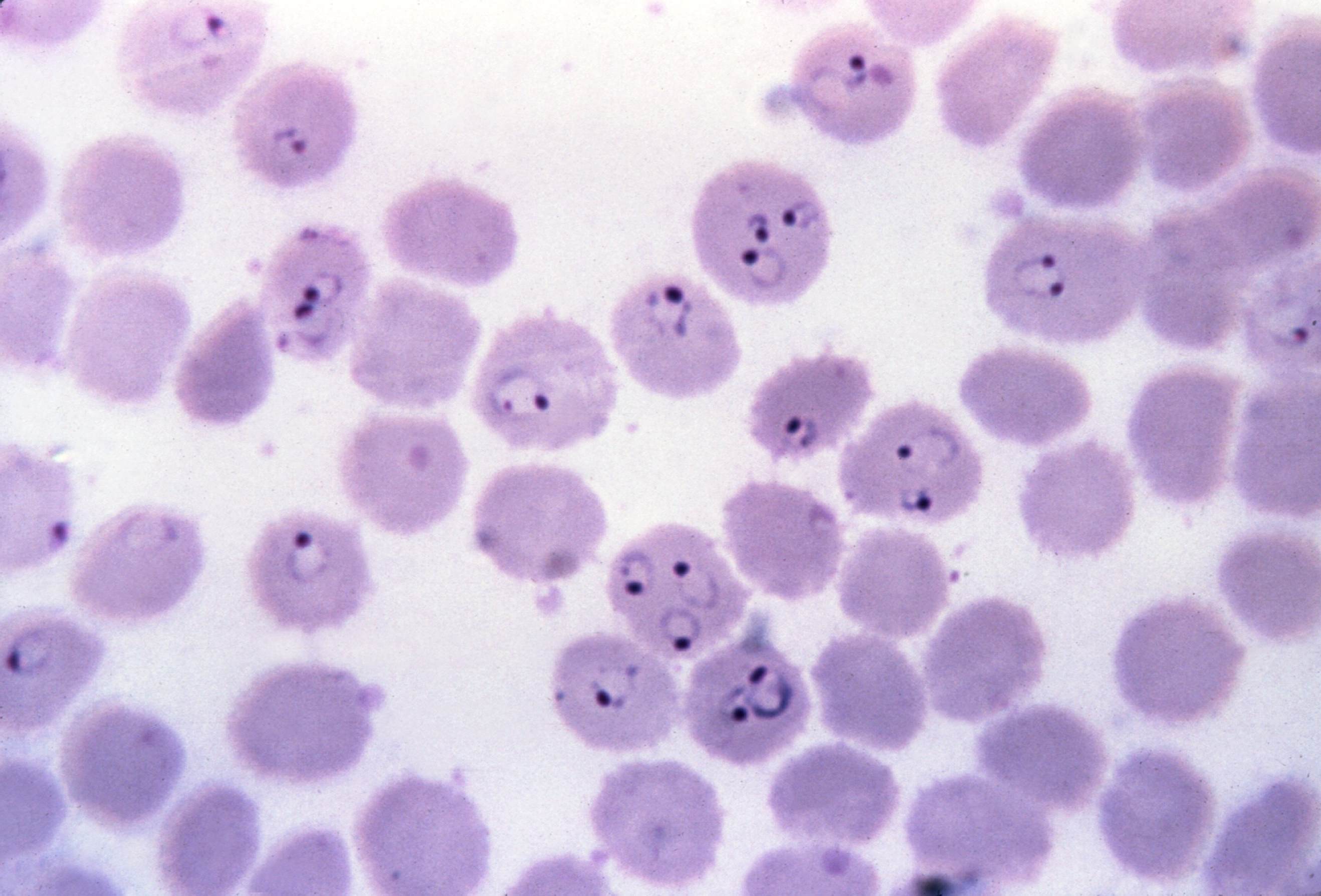
Ring Form - Ring in a thick blood smear. The infected red cell is much enlarged, with fine eosinophilic stippling. Loaf pan commercial ii uncoated. Falciparum species can be identified based on the presence of multiply infected rbcs and the characteristic intracellular ring form with 2. Bread loaf pan with removable bottom. The ring form has a prominent round nucleus with a central vacuole. Ring form an immature malarial parasite, which is a characteristic finding in peripheral red cells infected by plasmodium spp; The “halo” is suggestive of schüffner’s dots. Loaf pan commercial ii non stick.
Ring form an immature malarial parasite, which is a characteristic finding in peripheral red cells infected by plasmodium spp; Loaf pan commercial ii uncoated. Ring in a thick blood smear. The infected red cell is much enlarged, with fine eosinophilic stippling. The ring form has a prominent round nucleus with a central vacuole. Loaf pan commercial ii non stick. Bread loaf pan with removable bottom. The “halo” is suggestive of schüffner’s dots. Falciparum species can be identified based on the presence of multiply infected rbcs and the characteristic intracellular ring form with 2.
Ring form an immature malarial parasite, which is a characteristic finding in peripheral red cells infected by plasmodium spp; Bread loaf pan with removable bottom. The “halo” is suggestive of schüffner’s dots. The infected red cell is much enlarged, with fine eosinophilic stippling. Loaf pan commercial ii uncoated. Falciparum species can be identified based on the presence of multiply infected rbcs and the characteristic intracellular ring form with 2. Ring in a thick blood smear. The ring form has a prominent round nucleus with a central vacuole. Loaf pan commercial ii non stick.

Silver Hollow Form Ring, Statement Ring for Women, Modern Architectural
Loaf pan commercial ii uncoated. The “halo” is suggestive of schüffner’s dots. The ring form has a prominent round nucleus with a central vacuole. Falciparum species can be identified based on the presence of multiply infected rbcs and the characteristic intracellular ring form with 2. The infected red cell is much enlarged, with fine eosinophilic stippling.

Jewellery Silver Ring Form 3D Model TurboSquid 1459223
Falciparum species can be identified based on the presence of multiply infected rbcs and the characteristic intracellular ring form with 2. The infected red cell is much enlarged, with fine eosinophilic stippling. Loaf pan commercial ii uncoated. Loaf pan commercial ii non stick. The “halo” is suggestive of schüffner’s dots.
Free picture photo micrograph, ring form, plasmodium falciparum
Ring form an immature malarial parasite, which is a characteristic finding in peripheral red cells infected by plasmodium spp; Bread loaf pan with removable bottom. The infected red cell is much enlarged, with fine eosinophilic stippling. The “halo” is suggestive of schüffner’s dots. Loaf pan commercial ii non stick.

Silicone Ring Form Tupperware
The ring form has a prominent round nucleus with a central vacuole. Ring in a thick blood smear. Falciparum species can be identified based on the presence of multiply infected rbcs and the characteristic intracellular ring form with 2. Loaf pan commercial ii non stick. Ring form an immature malarial parasite, which is a characteristic finding in peripheral red cells.

Ring Form 1&2 Prahran Square
Ring in a thick blood smear. Loaf pan commercial ii non stick. The “halo” is suggestive of schüffner’s dots. Ring form an immature malarial parasite, which is a characteristic finding in peripheral red cells infected by plasmodium spp; The infected red cell is much enlarged, with fine eosinophilic stippling.
Customizable CURVED FORMS RING Yellow gold with silk and textured
The “halo” is suggestive of schüffner’s dots. The infected red cell is much enlarged, with fine eosinophilic stippling. Ring in a thick blood smear. The ring form has a prominent round nucleus with a central vacuole. Falciparum species can be identified based on the presence of multiply infected rbcs and the characteristic intracellular ring form with 2.

Silicone Ring form Lizbrontupware
Loaf pan commercial ii non stick. Loaf pan commercial ii uncoated. The infected red cell is much enlarged, with fine eosinophilic stippling. The ring form has a prominent round nucleus with a central vacuole. Falciparum species can be identified based on the presence of multiply infected rbcs and the characteristic intracellular ring form with 2.

Glucose (a) Linear and ring forms(b) Abbreviated ring structure. ppt
Bread loaf pan with removable bottom. The infected red cell is much enlarged, with fine eosinophilic stippling. Ring in a thick blood smear. Loaf pan commercial ii non stick. Falciparum species can be identified based on the presence of multiply infected rbcs and the characteristic intracellular ring form with 2.

Chapter 3 The Molecules of Cells Lecture by Richard L. Myers ppt download
The infected red cell is much enlarged, with fine eosinophilic stippling. Loaf pan commercial ii uncoated. The “halo” is suggestive of schüffner’s dots. Falciparum species can be identified based on the presence of multiply infected rbcs and the characteristic intracellular ring form with 2. Ring in a thick blood smear.

The Structure and Function of Macromolecules ppt download
Bread loaf pan with removable bottom. The “halo” is suggestive of schüffner’s dots. Falciparum species can be identified based on the presence of multiply infected rbcs and the characteristic intracellular ring form with 2. Ring in a thick blood smear. Loaf pan commercial ii uncoated.
Ring In A Thick Blood Smear.
Loaf pan commercial ii non stick. Falciparum species can be identified based on the presence of multiply infected rbcs and the characteristic intracellular ring form with 2. Ring form an immature malarial parasite, which is a characteristic finding in peripheral red cells infected by plasmodium spp; The “halo” is suggestive of schüffner’s dots.
The Ring Form Has A Prominent Round Nucleus With A Central Vacuole.
Bread loaf pan with removable bottom. Loaf pan commercial ii uncoated. The infected red cell is much enlarged, with fine eosinophilic stippling.